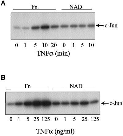
Figure 7.
Activation of JNK is anchorage dependent in HUVECs. Confluent HUVECs were serum starved, trypsinized, and suspended in medium containing 2% BSA. Following a 1-h period in suspension, cells were allowed to adhere to dishes coated with either fibronectin (Fn) or held in suspension (NAD) for 3 h prior to agonist stimulation. (A) TNFα time course. Following a 3-h period of cells replated on fibronectin or held in suspension, cells were treated with TNFα for various times (0–20 min). (B) TNFα dose response. Cells were stimulated with TNFα (0–125 ng/ml) for 10 min. Cells were lysed, and cell lysates were analyzed using an in vitro JNK assay, as described in MATERIALS AND METHODS. The autoradiograms represent the 32P phosphorylation of the substrate c-Jun.

