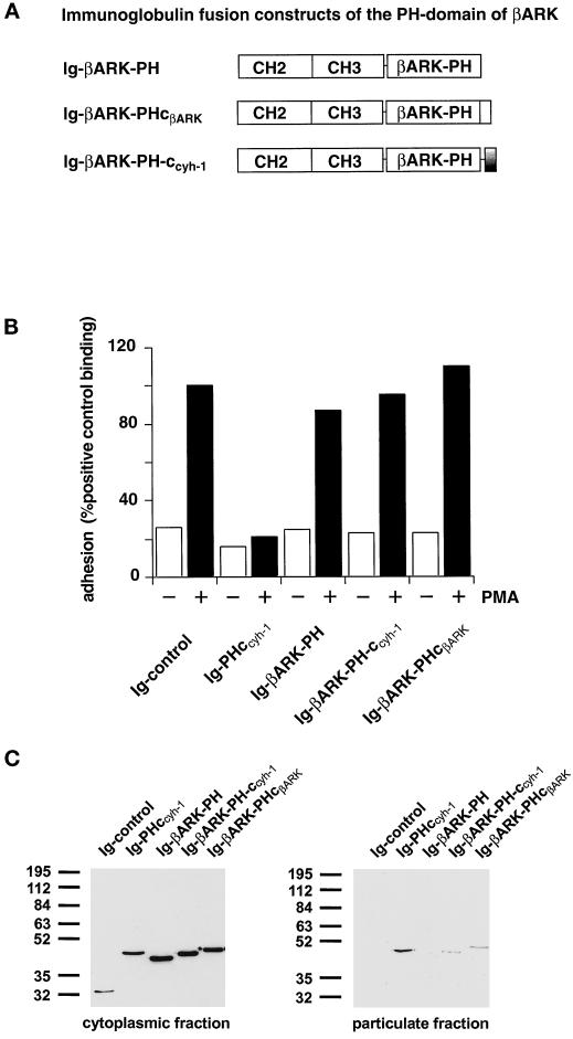
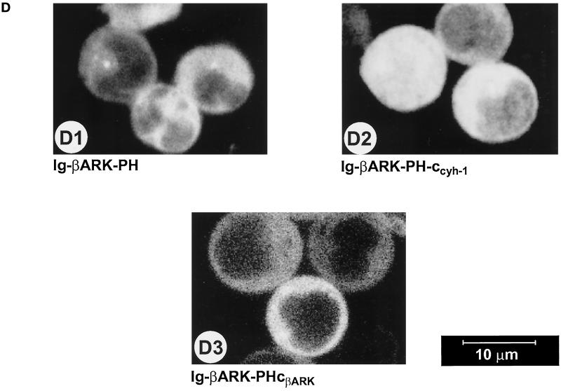
Figure 5.
(A) Outline of the βARK-PH domain constructs used in this part of the study. In the Ig-βARK-PH-ccyh-1 construct, the c domain of cytohesin-1 has been fused to the PH domain of βARK. (B) Adhesion assay of βARK-PH fusion proteins. Neither the wild-type βARK-PH domain and polybasic c-terminus nor a fusion protein of the βARK-PH domain with the c domain of cytohesin-1 (Ig-βARK-PH-ccyh-1) interferes with induced adhesion of Jurkat cells to ICAM-1. (C) Cellular fractionation of βARK-PH fusion proteins. Polybasic elements of either βARK or cytohesin-1 do not cooperate significantly with the βARK-PH domain in membrane association. (D facing page) Confocal laser scans of the subcellular distribution of βARK-PH fusion proteins. All tested fusion proteins are expressed in the cytoplasm.


