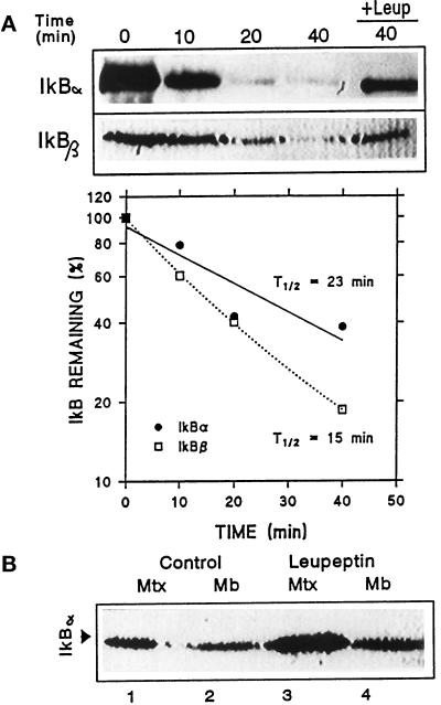
Figure 2.
Degradation of IκB inside lysosomes. (A) Rat liver lysosomes were incubated in an isotonic medium at 37°C in the absence or presence (+Leup) of leupeptin. At the indicated times, reactions were stopped by addition of Laemmli sample buffer and levels of IκB α and β present in the lysosomal fraction were detected by immunoblot following SDS-PAGE. The graph shows the quantification of four different experiments similar to the one shown and the calculated half-life for both proteins. (B) Lysosomes isolated from liver of nontreated or leupeptin-treated rats (see MATERIALS AND METHODS) were subjected to hypotonic shock and matrix and membranes were separated. Levels of IκB in each lysosomal fraction were detected by immunoblot with a specific antibody following SDS-PAGE.

