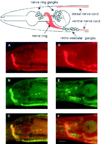
Figure 5.
Subcellular localization of UNC-11 in wild-type and unc-104 mutant nematodes. Confocal images of freeze-cracked nematodes double stained with affinity-purified rabbit α-UNC-11 antisera (first row); affinity purified mouse α-synaptotagmin antisera (second row), and merge (third row). (A–C) Wild-type. (D–F) unc-104(rh43). A diagram depicting the organization of nervous tissue in the C. elegans head including the position of neuronal cell bodies, the nerve ring neuropil, and the ventral and dorsal nerve cords is positioned above the confocal images. Synaptic-rich regions are depicted in red. Commissures, dendrites, minor process bundles, and many neuronal cell bodies of the ganglia were omitted for clarity. Bar, 10 μm.

