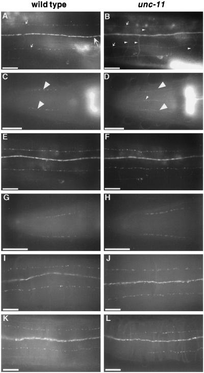
Figure 7.
Synaptobrevin, but not other synaptic vesicle proteins, is mislocalized in unc-11 mutants. The localization of vesicle proteins was examined in the wild type (first column) and in unc-11 animals (second column) using both GFP-tagged proteins and antibodies. (A and B) Images of the dorsal nerve cord of live immobilized adult animals expressing GFP-tagged synaptobrevin. The dorsal cord (large arrow), dorsal sublateral processes (small arrows), and commissural processes of motor neurons (small arrowheads) are labeled. The out-of-focus fluorescence in the lower right of each panel derives from the spermatheca that expresses synaptobrevin. (A) A wild-type animal. (B) unc-11(e47) animal. (C and D) Images of heads of adult worms fixed and stained with α-synaptobrevin antibodies and visualized with FITC-conjugated secondary antibodies. Synaptic varicosities of the SAB motor neuron processes are labeled with large arrowheads in a wild-type animal (C) and an unc-11(e47) animal (D). Note the appearance of synaptobrevin immunoreactivity in ventral processes (probably AVM and VA1) in panel D. (E and F) Images of the dorsal nerve cord of live immobilized adult animals expressing GFP-tagged synaptogyrin in a wild-type animal (E) and a unc-11(e47) animal (F). (G–J) Images of adult worms fixed andstained with α-RAB-3 antibodies and visualized with FITC-conjugated secondary antibodies in the head of a wild-type animal (G), and an unc-11(e47) animal (H); and a portion of the dorsal cord of a wild-type animal (I) and an unc-11(e47) animal (J). (K and L) Images of a portion of the dorsal cord of adult hermaphrodite worms fixed and stained with α-synaptotagmin antibodies and visualized with FITC-conjugated secondary antibodies in a wild-type animal (K) and an unc-11(n2954) animal (L). Anterior is left in all images. Bar, 10 μm.

