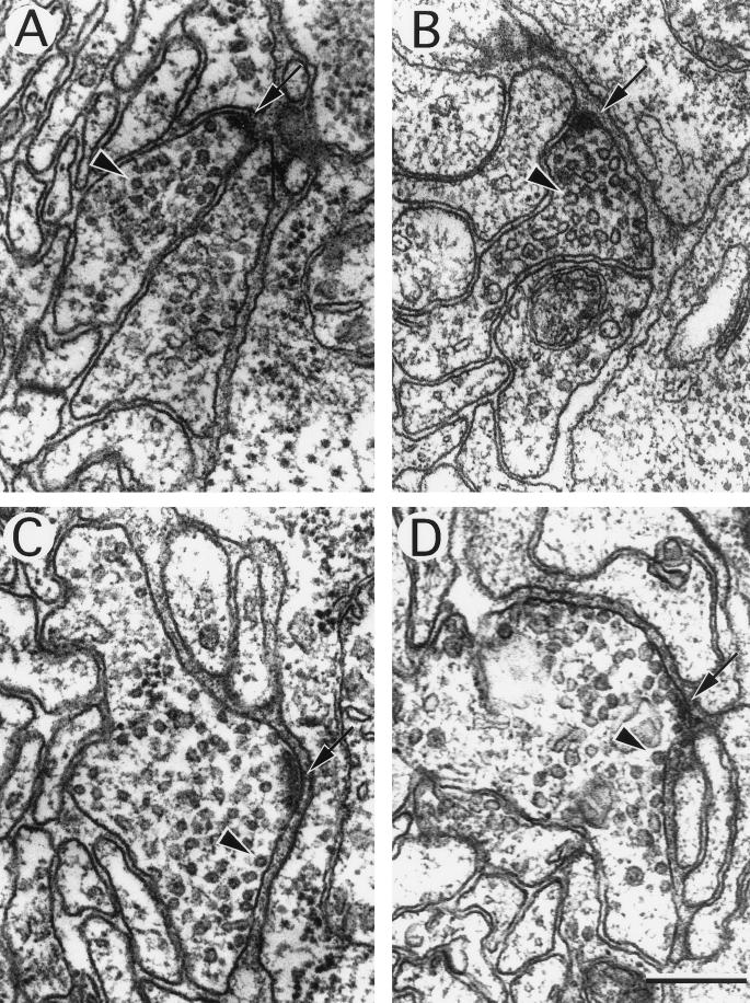
Figure 9.
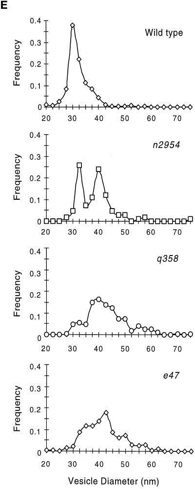
Ultrastructure of neuromuscular junctions from wild-type and unc-11 mutants. Electron micrographs of the ventral nerve cord of young adult animals. The neuronal cell type was deduced from examination of serial sections. A thick electron-dense presynaptic specialization is visible at the apposition of nerve and muscle in each image (arrow), and a single synaptic vesicle is indicated (arrowhead) in each micrograph. (A) Cholinergic neuromuscular junction in a wild-type animal. (B) Cholinergic neuromuscular junction in an unc-11(q358) mutant. (C) GABAergic neuromuscular junctions in a wild-type animal. (D) GABAergic neuromuscular junction in an unc-11(e47) mutant. Note the increase in vesicles in contact with the membrane adjacent to the postsynaptic muscle. (E) Quantitation of vesicle diameter from wild-type and unc-11 neuromuscular junctions. Vesicle diameters were measured in sections containing active zones and binned into 2.5-nm intervals. The fraction of vesicles in each size class is plotted at the midpoint of each binning interval (wild-type, n = 305; unc-11(e47), n = 214; unc-11(q358), n = 197; unc-11(n2954), n = 109). Bar, 200 nm.


