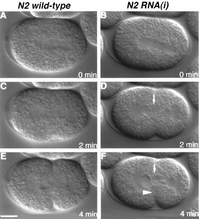
Figure 6.
Time-lapse Nomarski images of first cleavage. The embryo in the left column is wt; the embryo in the right column is the offspring of an animal injected with antisense zen-4 RNA. Anterior is to the left in all panels. (A) wt Metaphase embryo. (B) Metaphase embryo lacking ZEN-4. At this time, the spindle appears normal. (C) wt Anaphase embryo. Initiation of the cleavage furrow alters the ellipsoid shape of the embryo. (D) The cleavage furrow resembles the wt furrow (C), demonstrating that furrow initiation does not require zen-4. The white arrow points to the cytoplasmic granules in the spindle midzone, suggesting a lack of organized midzone microtubules. (E) wt Telophase embryo. The cleavage furrow is approaching the spindle midzone. (F) The white arrowhead points to the limit of furrow progression in an embryo lacking ZEN-4. Scale bar, 10 μm.

