Figure 3.
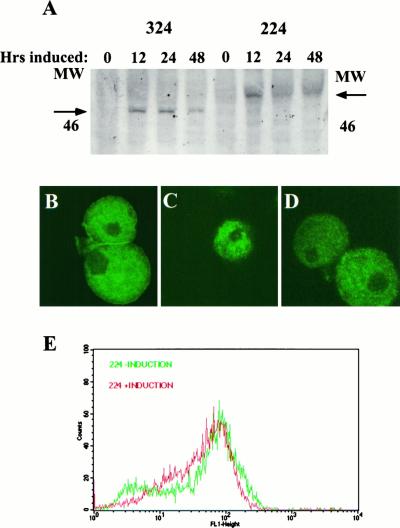
Inducible expression of the 224 and 324 fusion proteins in E. histolytica. (A) Time course of expression of lectin–GFP fusion proteins after induction with tetracycline. Stably transfected pGIR324/308 and pGIR224/308 amebae were induced to express the 324 and 224 fusion proteins, respectively, with tetracycline for the indicated times. Amebic proteins were analyzed by SDS-PAGE followed by Western blotting with anti-GFP antibodies. The positions of the 324 and 224 fusion proteins and the 46-kDa size marker are shown. (B–D) Intracellular location of lectin–GFP fusion proteins. Control amebae (B) and amebae induced to express the 224 (C) or 324 (D) fusion proteins were fixed in paraformaldehyde, permeabilized, and immunostained with polyclonal antibody to the lectin (B) or to GFP (C and D). Amebae were then incubated with FITC-conjugated sheep anti-rabbit IgG, mounted, and visualized by confocal microscopy. (E) Surface expression of the endogenous lectin is not altered by expression of the 224 lectin–GFP fusion protein. Amebae were incubated with antilectin antibody. After incubation with an FITC-conjugated secondary antibody, amebae (uninduced or induced to express 224 for 24 h) were analyzed by single parameter FACS for surface fluorescence.

