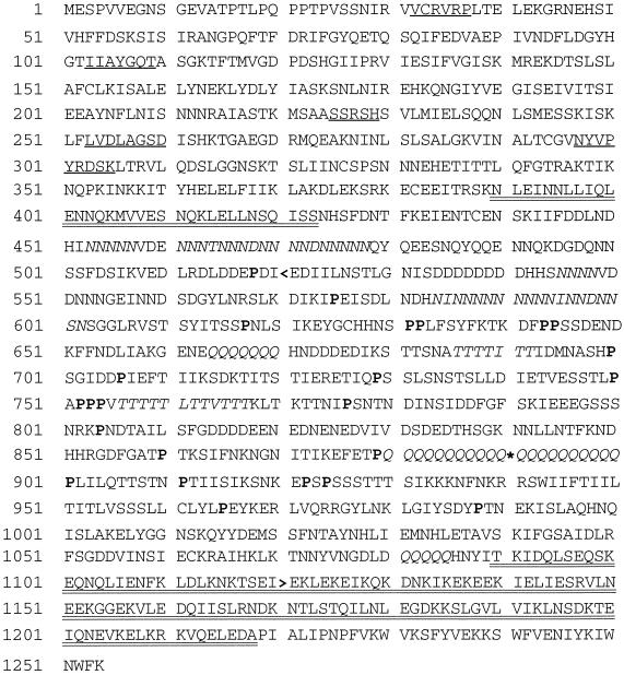
Figure 2.
Derived polypeptide sequence of K7. An asterisk indicates the position in the original clone corresponding to a breakpoint between the K7 cDNA and an extraneous sequence. Four conserved kinesin nucleotide binding loops and one microtubule binding loop (NYVPYRDSK) are underlined. Regions of the polypeptide sequence predicted to form a coiled-coiled structure are double underlined. Clusters of Asn, Glu, and Thr residues are shown in italics. Pro residues in the central nonhelical core are shown in bold. A back arrowhead indicates the C terminus of the motor domain fragment used for immunization and in vitro motility studies.

