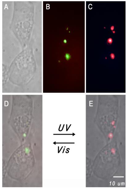
Figure 3.

Dual color fluorescent nanoparticles were transported into HEK-293 cells using liposomes as delivery vehicles; these internalized polymer nanoparticles can be selectively highlighted with either green or red fluorescence. (A) White light image of the cells under studies. Fluorescence imaging of the same cells is shown when the nanoparticles emit green (B) or red color (C), respectively. Images (D) and (E) are overlays of (A) and (B) or (A) and (C), respectively. A short UV pulse switches the highlighted green spots (D) to vivid red fluorescence (E) while visible light reverses the process.
