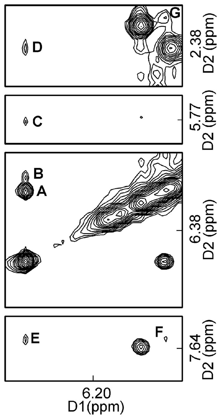
Figure 3.

1H NOESY spectrum showing the assignment of the exocyclic 1,N2-εdG protons H6 and H7, and NOEs between the etheno and DNA protons. The assignments are A, X6 H7→X6 H6; B, X6 H7→C18 H1′; C, X6 H7→C19 H5; D, X6 H7→C18 H2″; E, X6 H7→C19 H6; F, X6 H6→C19 H6; and G, X6 H6→C19 H2″. The spectrum was recorded in 10 mM NaH2PO4, 100 mM NaCl, 5 μM Na2EDTA, at 7 °C (pH 8.6).
