Figure 2.
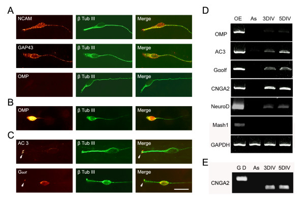
Cultured olfactory sensory neurons express characteristic markers. A) Cultured olfactory sensory neurons identified by β-tubulin III (β-Tub III) expression (green) also express neuronal cell adhesion molecule (NCAM; red) and GAP-43 (red) and olfactory marker protein (OMP; red) at 3 DIV. B) OMP positive cells were identified at 8 days in vitro (DIV). C) Adenylyl cyclase (AC)3 (arrowhead) and Gαolf (arrowhead and cell body) expression (red) at 5 DIV. D) OMP, AC3, Gαolf, cyclic nucleotide gated channel Ay subunit (CNGA2) and NeuroD expression were detected by RT-PCR at 3 DIV and 5 DIV. Mash1 expression was not detected at 3 DIV or 5 DIV. GAPDH expression served as quantity control. Olfactory epithelium (OE) cDNA was used as a positive control and feeder layer astrocyte (As) cDNA was used as a negative control. E) CNGA2 primers were designed to span an intron and, therefore, were able to discriminate PCR products amplified from genomic DNA (G D), which is 710 bp, or olfactory sensory neuron culture cDNA, which is 429 bp. Bar = 20 μm.

