Figure 4.
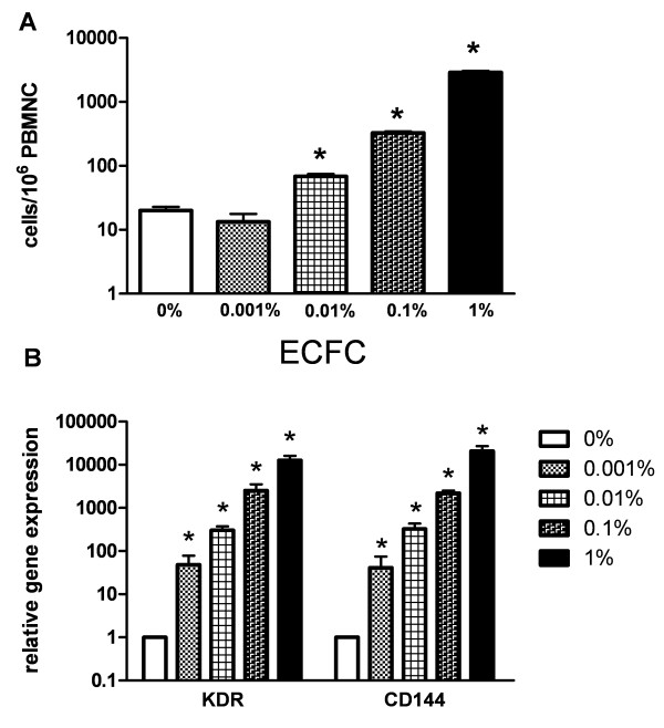
Detection of ECFC in peripheral blood samples. A) Four-channel flow cytometric analysis of autologous ECFC spiked into PBMNC of the respective donor (n = 5). ECFC were spiked at frequencies ranging from 0.001 to 1%. (* p < 0.05 compared to unspiked controls). B) Quantitative PCR analysis of ECFC marker gene expression in peripheral blood samples containing ECFC spiked at varying frequencies (0.001–1%; five independent experiments involving five different healthy donors). Expression of KDR and CD144 was analyzed in all samples relative to unspiked PBMNC. (* p < 0.05 compared to unspiked controls).

