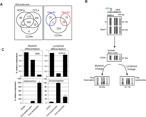
Figure 4. Cancer genes hypermethylated in somatic stem cells.
(A) The left-hand panel shows the numbers of sequences that are hypermethylated in the somatic stem cells CD34+, and hypermethylated in hESCs and CCLs. Note that most of the sequences hypermethylated in somatic stem cells are also hypermethylated in embryonic stem cells. The right-hand panel shows the number of sequences hypermethylated in CD34+ cells (black circle) classified as Class B-II genes (red circle). Sequences hypermethylated in CD34+ cells were never classified as Class B-I genes (blue circle). (B) Bisulfite genomic sequencing of multiple clones of the AIM2 promoter in Shef-1 and I3 stem cell lines (upper), CD34+ hematopoietic stem cell progenitors (middle), and terminally differentiated hematopoietic cells (peripheral lymphocytes and neutrophils). The color code is as for Figure 2. (C) Relationship between AIM2 and RUNX3 promoter hypermethylation and expression in CD34+ somatic hematopoietic stem cell progenitors and terminally differentiated hematopoietic cells (peripheral lymphocytes and neutrophils). The upper panel shows the relative methylation signal obtained with the methylation arrays and the lower panel the expression levels of AIM2 and RUNX3 mRNAs relative to GAPDH.

