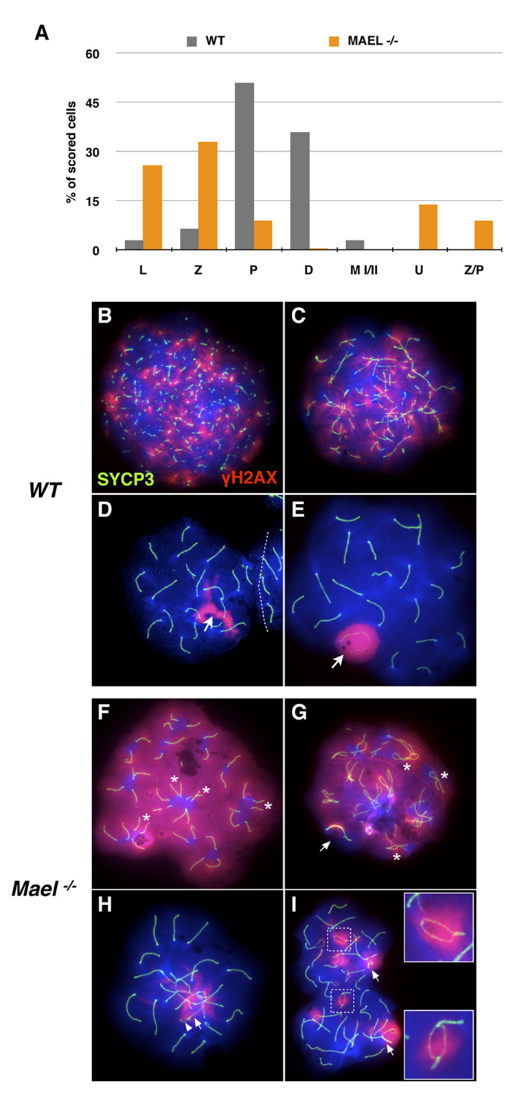
Figure 4. MAEL is required for chromosome synapsis.
Meiotic spreads in (B – I) are stained with antibodies against H2AX phosphorylated on S139 histone (red) and SYCP3 (green). DAPI labels DNA blue.
(A) Distribution of wild-type (gray bars, 400 scored nuclei) and Mael−/− (orange bars, 800 scored nuclei) spermatocytes among different substages of the meiotic prophase I (L - leptotene, Z - zygotene, P - pachytene, D - diplotene), metaphase (M I/II) and atypical classes (U − 40 univalents, Z/P - nuclei combining zygotene and pachytene features) of spermatocytes.
(B–E) Leptotene (B), zygotene (C), zygotene-pachytene transition (D) and pachytene (E) stages of the first meiotic prophase in wild type spermatogenesis.
(F–I) Representative examples of univalent (F), Z/P (G, H) and pachytene (I) Mael−/− spermatocytes.
(F) A Mael−/− spermatocyte with 40 univalent (fully asynaptic) chromosomes and high levels of DNA DSBs as indicated by γH2AX signal. Pairs of equal-sized axial elements in proximity of each other (asterisk) suggest homolog recognition.
(G) A Z/P Mael−/− spermatocyte with synapsed sex chromosomes (arrow) but only limited (asterisk) synapsis of autosomes and extensive DNA damage (γH2AX signal).
(H) A Z/P Mael−/− spermatocyte with minor autosomal asynapsis and synapsed X (arrow) and Y (arrowhead) chromosomes.
(I) A pachytene Mael−/− spermatocyte with asynapsed regions within bivalents (insets) and non-homologous synapsis (arrows).

