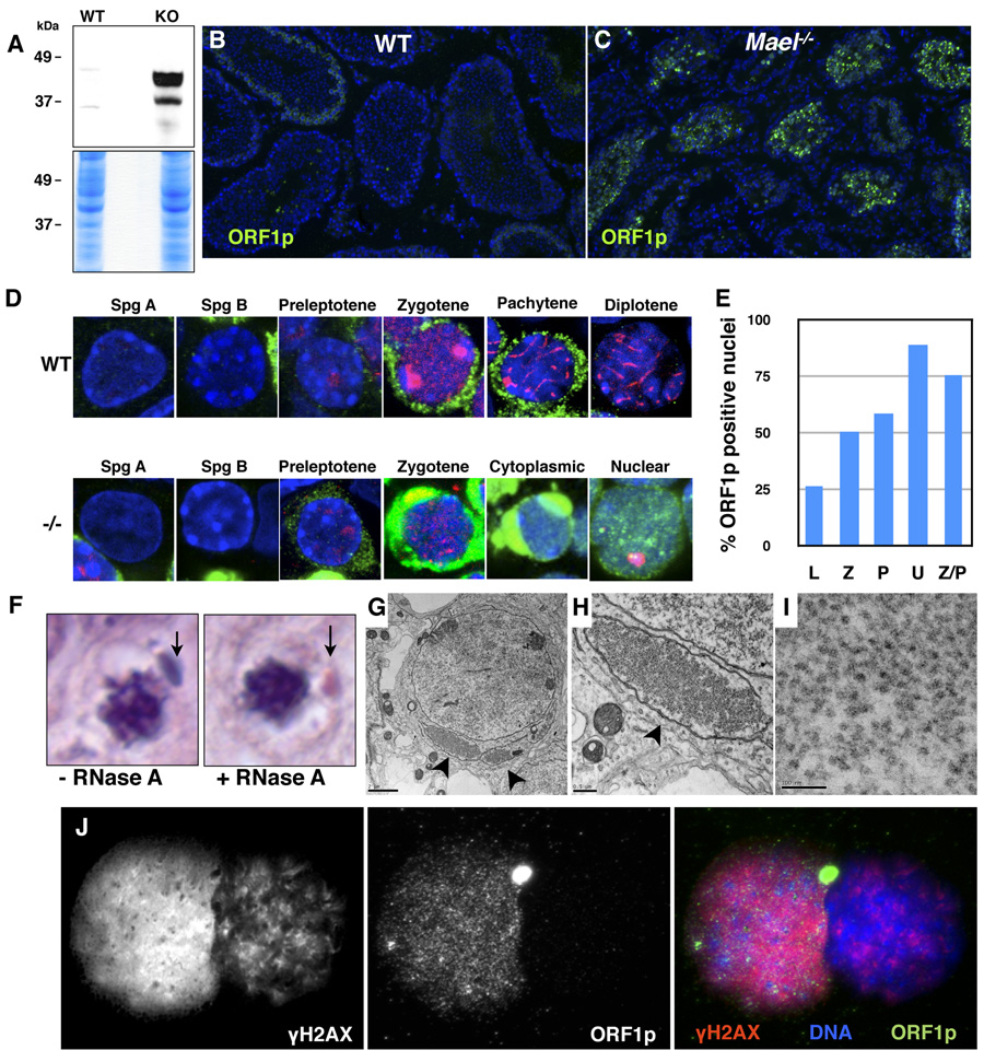
Figure 6. L1 ORF1p accumulation in cytoplasmic enclaves and nuclei of Mael−/− spermatocytes.
(A) Western blot analysis using an antibody to L1 ORF1p reveals a large increase in the protein levels in Mael−/− testis.
(B) Immunofluorescence using anti-ORF1p antibody on frozen sections of wild-type testes.
(C) Immunofluorescence using anti-ORF1p antibody on frozen sections of Mael−/− testes. A significant number of Mael−/− tubules contain spermatocytes with high levels of ORF1p in the cytoplasm and nuclei.
(D) ORF1p localization in wild type (WT) and Mael−/− (−/−) germ cells. Spg A – type A spermatogonia, Spg B – type B spermatogonia, Cytoplasmic – localization of ORF1p to cytoplasmic enclaves. Green - ORF1p, Red - SYCP3, DAPI stains DNA blue.
(E) Percent of ORF1p positive nuclei in various classes of Mael−/− spermatocytes described in Figure 4.
(F) Cytoplasmic enclaves (arrows) in a p18 Mael−/− spermatocytes. Shown are representative examples of spermatocytes from histological H & E sections either subjected or not to RNase A treatment prior to staining.
(G) Electron microscopy of Mael−/− testis reveals perinuclear domains (arrowheads) packed with presumed L1 RNPs (bar - 2 µm).
(H) A close-up view of the cytoplasmic enclave shown in (F) (bar − 0.5 µm).
(I) A close-up view of the presumed L1 RNPs in the cytoplasmic enclave (bar − 100 nm).
(J) Co-localization of ORF1p and γH2AX in the nucleus of a Univalent Mael−/− spermatocyte. γH2AX; ORF1p double positive spermatocyte is inferred to be a Univalent spermatocyte based on the pattern of γH2AX staining (high level in euchromatin, very little in heterochromatin; compare to Figure 4F).

