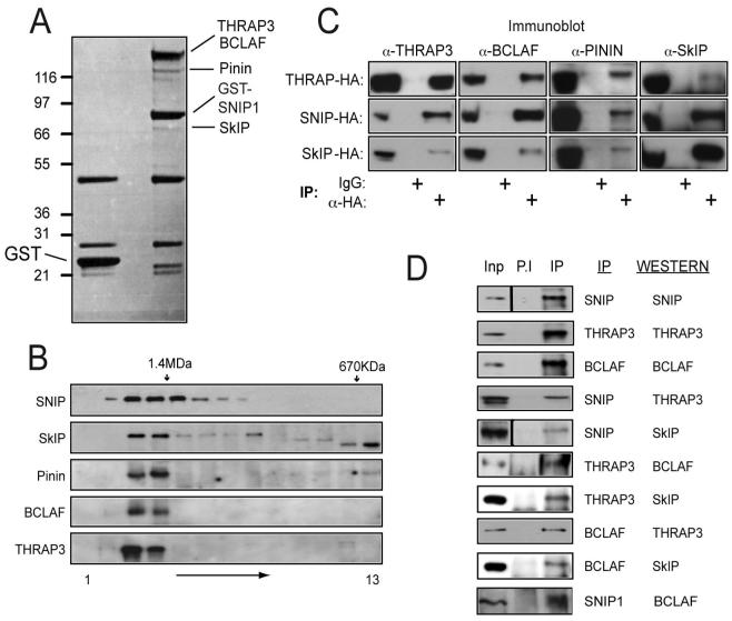
Figure 3. Identification of SNIP1 associated proteins.
(A) Purification of SNIP1 associated proteins. 10μg of expression plasmids encoding GST-SNIP1 or GST alone were transfected into HEK 293 cells and extracts were prepared prior to purification using glutathione agarose, SDS-PAGE and colloidal coomassie staining. Bands specifically present in the SNIP1-GST lane were excised and proteins identified by MALDI-Tof-Tof (MS/MS).
(B) SNIP1 associated proteins are all in a high molecular weight complex of similar size. HeLa nuclear extract was resolved by Superose 6 gel filtration and fractions separated by SDS-PAGE and western blotted using the antibodies indicated. The positions of molecular weight markers used during column calibration are shown.
(C) Confirmation of SNIP1 associated proteins by western blot analysis. 293 cells were transfected with THRAP3, SNIP1 or SkIP-HA expression vectors and anti-HA immunoprecipitations performed prior to SDS-PAGE and western blotting for endogenous proteins as indicated.
(D) Co-immunoprecipitation of endogenous SNIP1 associated proteins. 200μg of U-2 OS extract was analysed by immunoprecipitation, SDS-PAGE and western blotting using the indicated antibodies. Inputs represent 5% of total protein used in the assay.

