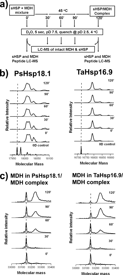
FIGURE 2.
Global HXMS of sHSP and substrate. a, experimental scheme for examining HX during sHSP-MDH complex formation. Concentrations of sHSP and MDH are as in Fig. 1. b, mass spectra of sHSP global exchange pattern as a function of time monitored by HXMS. Left, PsHsp18.1 in the presence of MDH. Three populations of PsHsp18.1 were present in the sample used in the experiment: unmodified protein, protein with N-terminal methylation and a very minor fraction of N-terminally acetylated protein. All forms bound substrate equally well (not shown). Right, TaHsp16.9 in the presence of MDH. Two populations of TaHsp16.9 were present in the protein used in the experiment: unmodified protein and a very minor fraction of N-terminally acetylated protein. c, mass spectra of MDH global HX pattern as a function of time. Left, MDH thermal unfolding in the presence of PsHsp18.1. Right, MDH thermal unfolding in the presence of TaHsp16.9.

