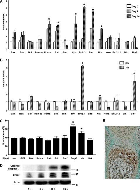
FIGURE 3.
Role of pro-apoptotic Bcl-2 family member proteins in Pi-induced cell death. A, expression patterns of pro-apoptotic Bcl-2 family member proteins in ATDC5 cells during ITS treatment as determined by time and RT-PCR. The results are expressed as the means ± S.D. of three cultures. Experiments were repeated at least three times, and the representative data are presented. *, significantly different from day 0, p < 0.05. B, expression patterns of pro-apoptotic Bcl-2 family member proteins in ATDC5 cells after Pi stimulation as determined by real time PCR. ATDC5 cells were pretreated with ITS for 14 days (0 h) before Pi stimulation (20 mm). The results are expressed as the means ± S.D. of three cultures. Experiments were repeated at least three times, and the representative data are presented. *, significantly different from time 0, p < 0.05. C, effect of gene silencing of pro-apoptotic Bcl-2 family member proteins on cell viability of primary chondrocytes 24 h after Pi stimulation. Gene silencing of Bnip3 and Nix significantly promoted the cell viability, p < 0.05. The results are expressed as the means ± S.D. of six cultures. Experiments were repeated at least three times, and the representative data are presented. GFP, green fluorescent protein. D, Western blot analysis after Pi stimulation. Protein levels of Bnip3 increased in ATDC5 cells after 6 h of Pi stimulation, which was followed by caspase-7 activation. Bnip3 was detected as two bands. E, immunohistological examination of a murine metatarsal bone (E18.5) with anti-Bnip3 antibody. Bnip3 expression was primarily localized in the hypertrophic layers of chondrocytes.

