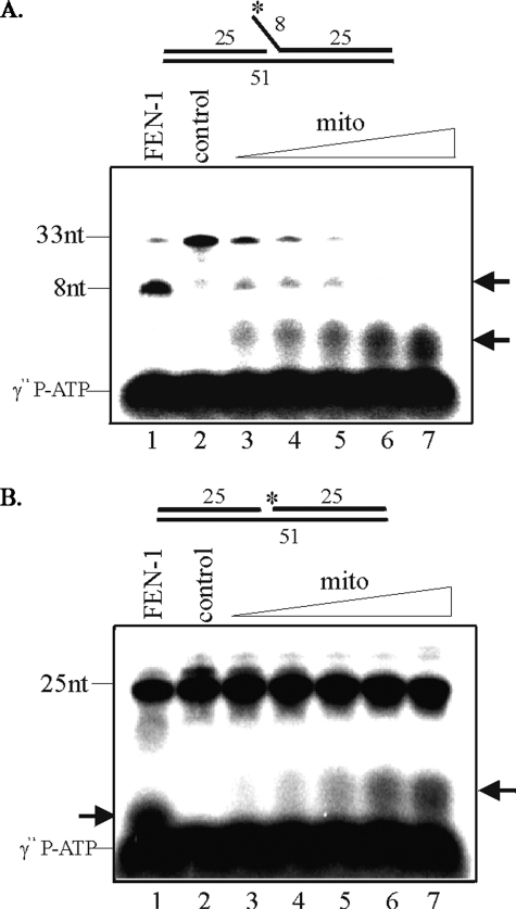
FIGURE 5.
Distinct mitochondrial 5′-exo/endonuclease activity in HCT116 cells. Substrates were labeled at the terminus of flap of oligo8 (A) or gap of oligo7 (B) as indicated by an asterisk, and increasing concentration of mitochondrial extracts was used in assays (lanes 3–7). Human recombinant FEN-1 was used as a positive control (lane 1). control, no extract.

