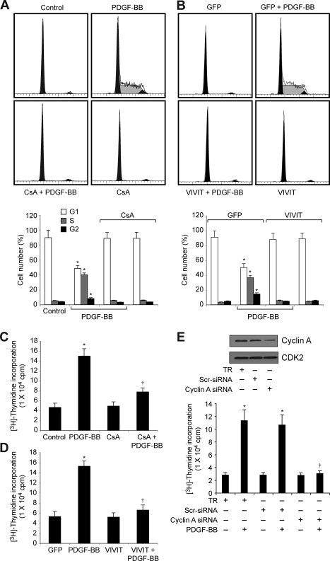
FIGURE 2.
Blockade of NFAT activation prevents cell cycle progression of VSMC from G1 to S phase. A, quiescent VSMC were treated with and without PDGF-BB (20 ng/ml) in the presence and absence of CsA (10 μm) for 16 h and subjected to FACS analysis. B, VSMC were transduced with either Ad-GFP or Ad-VIVIT at an m.o.i. of 80, quiesced, treated with and without PDGF-BB (20 ng/ml) for 16 h, and subjected to FACS analysis. Thebar graphs represent mean ± S.D. values of three independent experiments. C, quiescent VSMC were treated with and without PDGF-BB (20 ng/ml) in the presence and absence of CsA (10 μm) for 24 h, and DNA synthesis was measured by [3H]thymidine incorporation. D, conditions were the same as in C except that cells were transduced with either Ad-GFP or Ad-VIVIT at an m.o.i. of 80, and quiesced before subjecting to treatment with PDGF-BB and measuring DNA synthesis. E, top panel, VSMC were transfected with scrambled (Scr) or cyclin A siRNA, and 36 h later cell extracts were prepared, and an equal amount of protein from each condition was analyzed by Western blotting for cyclin A levels. Bottom panel, after transfection with scrambled or cyclin A siRNA, cells were quiesced and treated with and without PDGF-BB (20 ng/ml) for 24 h, and DNA synthesis was measured as described in C.*, p < 0.01 versus control, GFP, or TR; †, p < 0.01 versus PDGF-BB or GFP + PDGF-BB or TR + PDGF-BB treatment alone. TR, transfection reagent.

