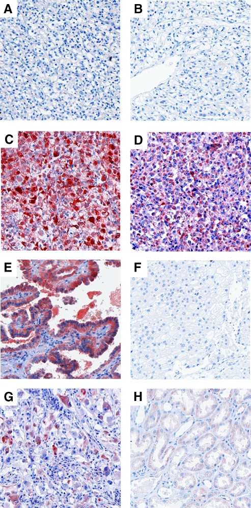
Figure 1.
Immunohistochemical detection of DcR3 protein in RCCs of diverse histopathologic subtypes (original magnification, x100). (A–D) Clear-cell (conventional) RCCs with different grades of malignancy (A: G1, B: G2, C: G3, D: G4); (E) papillary (chromophil) RCC; (F) chromophobe RCC; (G) collecting duct carcinoma; (H) normal tubular renal tissue. The IHSs were as follows: A, 0; B, 0; C, 12; D, 12; E, 12; F, 0; G, 9.

