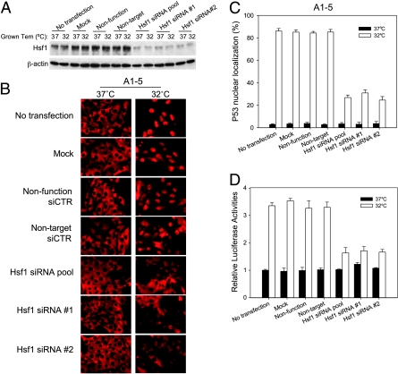
Figure 7.
Suppression of Hsf1 with siRNA inhibits p53 nuclear localization. (A) A1–5 cells were transfected with the indicated siRNA at 37°C. Forty-eight hours later, either the cells were harvested and extracts were prepared (37°C) or the incubation temperature was shifted to 32°C for 5 hours before preparing extracts (32°C). Levels of Hsf1 were determined by immunoblot analysis with the appropriate antibody. β-Actin levels were used as loading controls. (B) A1–5 cells were grown on coverslips at 37°C to 60% confluence and then transfected with the indicated siRNA. Forty-eight hours later, the coverslips were either immediately stained for p53 localization (37°C) or shifted to 32°C for 5 hours and then stained for p53 localization by PAb421. (C) The fraction of cells with nuclear localized p53 in panel (B) was quantified. Graphs show the mean ± SE from three independent experiments. (D) A1–5 cells stably transfected with pWWP-Luc-Neo plasmid were grown at 37°C to 60% confluence. Forty-eight hours after transfection with the indicated siRNA, either the cells were harvested immediately and assayed for luciferase activity (37°C) or the incubation temperature was shifted to 32°C for an additional 6 hours and then assayed for luciferase activity (32°C). Graphs show the mean ± SE from three independent experiments.

