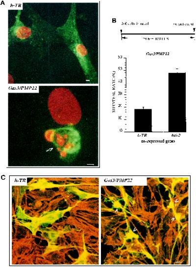
Figure 2.
gas3/PMP22-dependent biological activities in Schwann cells. (A) Confocal generated overlay showing nuclear morphology in rat Schwann cells overexpressing Gas3/PMP22 or h-TR. Schwann cells 24 h after seeding were microinjected with pGDSV7-gas3/PMP22 (100 ng/μl) or with pGDSV3-hTR (100 ng/μl); after 24 h cells were fixed and processed for immunofluorescence analysis to visualize Gas3/PMP22 or h-TR using the specific antibodies (green). Propidium iodide was used to visualize nuclei (red). Images were overlayered using a Zeiss confocal microscope and are displayed in pseudocolor. The arrow indicates a Gas3/PMP22-overexpressing cell. Bar, 5 μm. (B) bcl-2 and h-TR were coexpressed with gas3/PMP22 in Schwann cells. After 24 h from microinjection cells were fixed and processed for immunofluorescence to detect Gas3/PMP22. Survival was scored as described in the text. Data represent arithmetic means ± SD for five independent experiments (p < 0.001). (C) Confocal generated overlay showing actin architecture and cell morphology in Schwann cells cells coexpressing Gas3/PMP22 Bcl-2 and Gas2 or h-Tr Bcl-2 and Gas2. Schwann cells 24 h after seeding were microinjected with pGDSV3-hTR (50 ng/μl), pGDSV7-bcl-2 (50 ng/μl), and pGDSV7-gas2 (25 ng/μl) or with pGDSV7-gas3/PMP22 (50 ng/μl), pGDSV7-bcl-2 (50 ng/μl), and pGDSV7-gas2 (25 ng/μl). After 24 h cells were fixed and processed for immunofluorescence analysis to visualize Gas2 (green), using the specific antibody and actin filaments (red) and rhodamine-phalloidin. Images were overlayered using a Zeiss confocal microscope and are displayed in pseudocolor. Bar, 15 μm.

