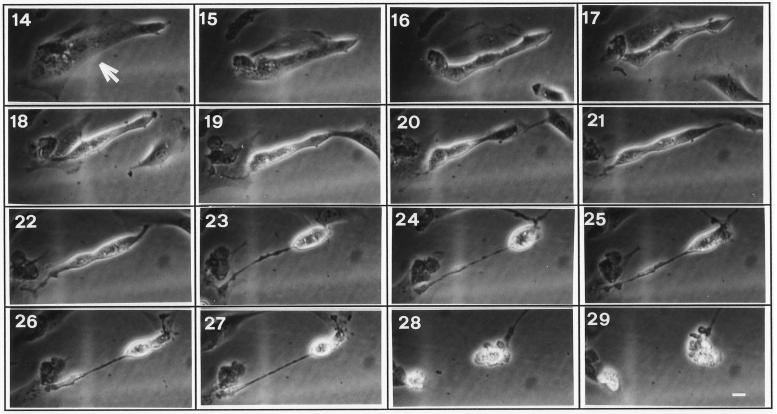
Figure 3.
Time-lapse images of a NIH3T3 cell overexpressing gas3/PMP22. Representative cell (arrow) injected with pGDSV7-gas3/PMP22 (100 ng/μl). Pictures at selected times after microinjection (as indicated) show the morphological changes from 17 h and the appearance of membrane blebbing at 28 h. Bar, 20 μm.

