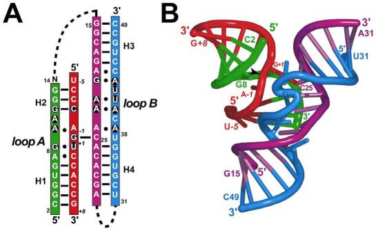
FIGURE 1.

Schematic diagrams of the minimal all-RNA hairpin ribozyme used in this and the previous study (ref. 11). (A) Secondary structure depiction based on the known structure. The substrate strand is red; other ribozyme strands are colored green, purple or blue. This color scheme is preserved throughout the figures. Lines indicate W-C H-bond pairs; filled circles represent non-canonical interactions. Dashed lines indicate covalent connections that were excluded from this construct. The loop A and B domains are labeled with conserved residues on black circles. Helices 1-4 are labeled (H1 to H4). Position 14 was omitted in this study. (B) Ribbon diagram of the refined 2.05 Å resolution G8 native hairpin ribozyme of this study, which is representative of the core fold. The scissile bond is indicated by an arrowhead. Positions A−1, G+1 and G8 are labeled and represent the active site.
