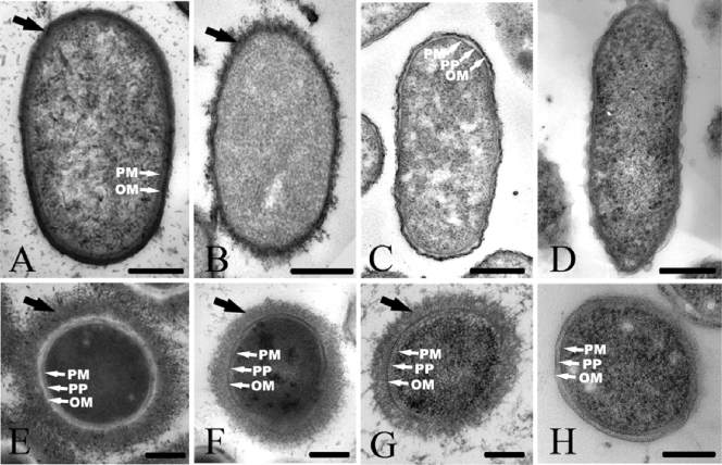
FIG. 1.
Representative TEM images of thin sections of E. coli K30 (A and E), P. aeruginosa FRD1 (B and F), S. oneidensis MR-4 (C and G), and G. sulfurreducens PCA (D and H). The samples were prepared by conventional embeddings with Ruthenium red staining (A to D) and freeze-substitution (E to H). The black arrows indicate the bacterial capsule, and the white arrows indicate the plasma membrane (PM), periplasm (PP), and outer membrane (OM). Bars, 250 nm.

