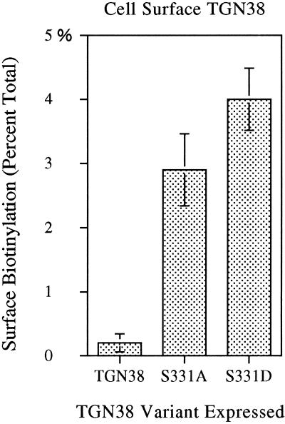
Figure 7.
Detection of cell-surface expression of TGN38 variants by surface biotinylation. Cells induced to comparable expression levels were cooled on ice and biotinylated with disulfide-linked biotin for 30 min to label cell surface proteins. Biotinylated proteins were affinity isolated from cell lysates with streptavidin-agarose beads. The remaining TGN38 was immunoprecipitated with a polyclonal antibody to TGN38 (shG29). Affinity- and immuno-precipitated proteins were processed and analyzed by densitometry as described in MATERIALS AND METHODS. Cell surface (biotinylated) TGN38 is expressed as the percentage of total TGN38 (biotinylated plus unbiotinylated) affinity isolated in each case (± SD, n = 3). (In parallel control samples, no endocytosis was observed in cells incubated on ice.)

