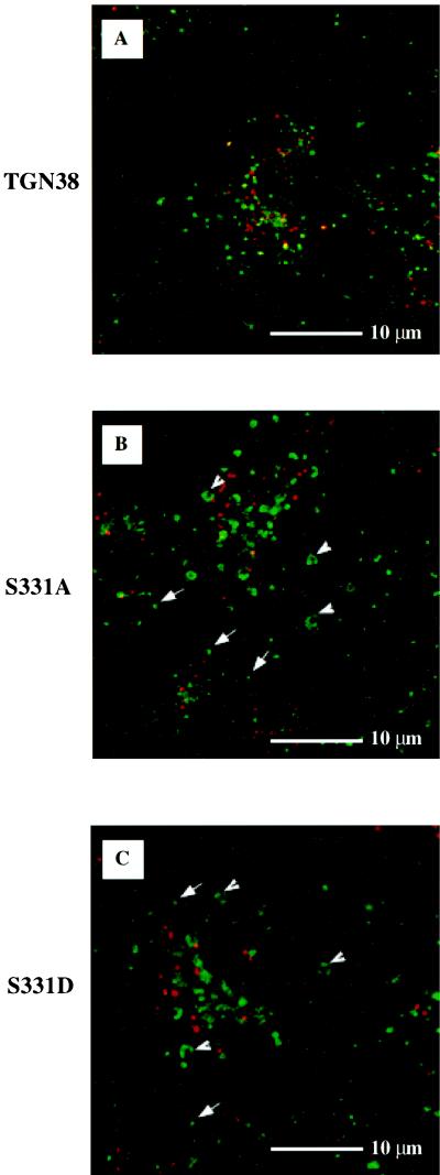
Figure 9.
Antibody uptake after a GPN-induced lysosomal block. Cells expressing wild-type TGN38 (A), S331A (B), or S331D (C) were preincubated for 10 min in the presence of 200 μM GPN to induce swollen, disfunctional lysosomal vacuoles (arrowheads). 2F7.1 ascites was then added to the tissue culture medium at a dilution of 1:400 in the continued presence of GPN. Concomitantly, Texas Red-conjugated transferrin was added at 10 μg/ml to label endosomal compartments. Cells were then incubated for 25 min at 37°C and processed for immunofluorescence using FITC-conjugated anti-mouse Ig to detect endocytosed 2F7.1. Arrows, nonswollen transport vesicles.

