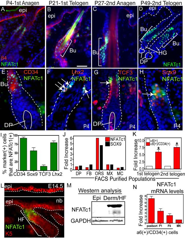
Figure 1. NFATc1: a marker of hair follicle stem cells.
(A-D) NFATc1 is expressed the upper HF during anagen at P4 and P27 and at the base of the follicle during telogen (P21 and P49). β4 integrin marks the dermo-epidermal interface. (C-H) Immunohistochemistry showing NFATc1 colocalization with bulge cell markers, CD34, Lhx2, TCF3 and Sox9. Arrows denote examples of co-expression. (I) Quantification of the % of cells with bulge cell markers that colocalize with NFATc1. Data are mean ± SEM. N=50−176 cells. (J-K) Real-time PCR analysis of NFATc1 and Sox9 mRNA in FACS isolated populations at P4 (J) and NFATc1 mRNA in the α6/CD34-positive bulge compartment during the 1st (P19) and 2nd (P49) telogen. (K). Data are mean ± SEM. N=2 (J) and N=3 (K) FACS isolated populations. Data are mean ± SEM. Asterisks indicate significance, p<0.05. (L) Expression of NFATc1 in the keratin 5 (K5)(+) epidermis at E14.5 or newborn (nb). (M) Isolated P4 epidermis and the dermis (containing HFs) were subjected to western analysis. (N) Real Time PCR analysis of NFATc1 mRNA from FACS isolated α6(+)/CD34(+) bulge cells, after passage (P1, P4) and in epidermal keratinocytes (MK). Abbreviations: DP, dermal papillae; Bu, bulge; ORS, outer root sheath, epi, epidermis, derm, dermis HG, hair germ; FB, fibroblasts; MX, matrix, MC, melanocytes. Dapi staining (blue) shows nuclear localization. Scale Bars, 30 μm.

