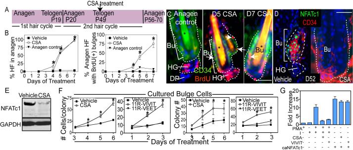
Figure 2. Impairing calcineurin/NFAT signaling results in hair growth and stem cell proliferation.
(A) Schematic illustrating the experimental design of CSA experiments. (B-C) Quantification of anagen induction and BrdU incorporation in follicle stem cells of the bulge (Bu) with cyclosporine A (CSA) treatment. The anagen control used was the first hair cycle (P18-P25). Data are the mean ± SEM for 50−100 follicles for 3 individual mice for each timepoint. Asterisks in (C) indicate hair shaft autofluorescence. (D-E) Immunohistochemistry and western analysis of NFATc1 expression following 3d treatment with vehicle or CSA. (F) Colony formation and cell number of bulge cells following treatment with vehicle, CSA, 11R-VIVIT, or 11R-VEET. N= 3 experiments with independent sorted populations. (G) NFAT reporter activity in response to calcium ionophore (I), phorbol myristate acetate (PMA), caNFATc1, CSA or 11R-VIVIT. Data are mean ± SEM. N=3−5 individual experiments for each treatment. Asterisks indicate significance, p<0.05. Scale bars, 30μm. Dapi staining (blue) shows nuclear localization. Abbreviations: HF, hair follicle; Bu, bulge; HG, hair germ; DP, dermal papillae.

