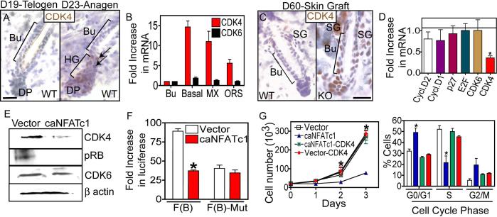
Figure 5. NFATc1 acts by repressing CDK4 expression to regulate G1/S progression in hair follicle stem cells.
(A) Anti-CDK4 immunohistochemistry during the telogen (D19) to anagen (D23) transition. Hematoxylin counterstain. (B) Real Time PCR analysis of CDK4 and CDK6 mRNA expression in bulge (Bu), all basal (α6+), ORS and matrix (MX) cell populations. Data are mean ± SEM. N=3 for each sample. (C) Immunostaining of CDK4 in WT and NFATc1-null follicles. Bu, bulge. (D) Expression of cell cycle regulatory genes in control or caNFATc1 expressing cells. Data are mean ± SEM. N=3 individual experiments. (E) Western analysis for CDK4, phosphorylated retinoblastoma protein (Rb), and CDK6 in control and caNFATc1 expressing keratinocytes (MKs). (F) Reporter assays using the CDK4 promoter with NFAT sites (F(B)) or mutated NFAT sites (F(B)-mut) in conjuction with caNFATc1 in MKs. Data are mean ± SEM. N=4 individual experiments. (G-H) Expression of CDK4 rescues MK growth and S phase progression of cells expressing caNFATc1. N=3 individual experiments. Data are mean ± SEM. Asterisks indicate significance, p<0.05. Scale Bar, 30 μm.

