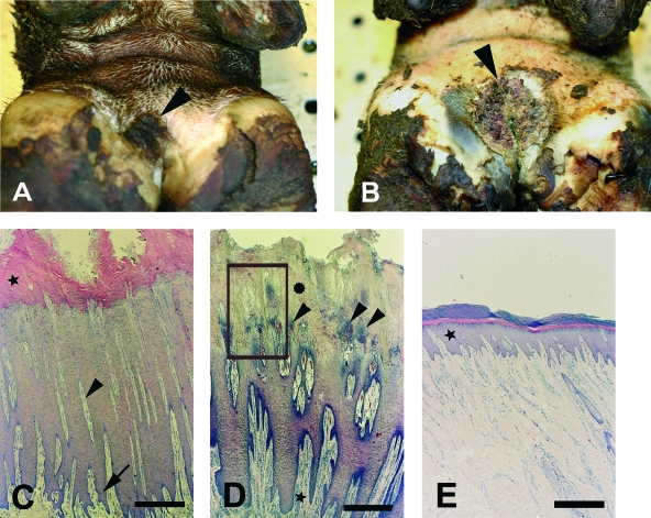
FIG. 1.
Gross histopathology of bovine DD in the interdigital area. (A) Biopsy specimen no. 40, showing circumscribed, focal, moist plaque with increased thickness of epidermis (arrowhead). (B) Biopsy specimen no. 41, showing circumscribed, ulcerated lesion with papillomatous proliferations (arrowhead). (C) Cross-section of biopsy specimen no. 30, revealing severe epidermal hyperplasia of the stratum spinosum with long, slender dermal papillae (arrowhead) and deep epidermal (arrow) and orthokeratotic (★) hyperkeratosis. Staining was done with H&E. Bar = 0.5 mm. (D) Cross-section of biopsy specimen no. 38, revealing severe epidermal hyperplasia of the stratum spinosum with bacterial colonization and exudation from dermal papillae (arrowhead). Keratinolysis of stratum corneum ( ) and perivascular dermatitis (★) are also shown. Staining was done with H&E the boxed area represents an area similar to that shown in Fig. 3). Bar = 0.5 mm. (E) Skin from the interdigital area, with a normally appearing epidermis (★). Staining was done with H&E. Bar = 0.5 mm.

