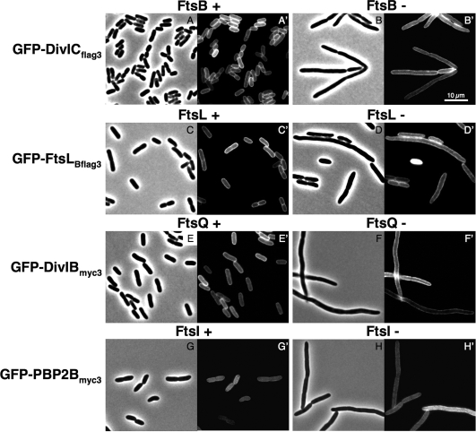
FIG. 2.
DivICflag3, FtsLBflag3, DivIBmyc3, and PBP 2Bmyc3 GFP fusions do not localize to the cell division site in strains depleted of their respective E. coli orthologs. E. coli strains were deleted of ftsB, ftsL, ftsQ, or ftsI at their native loci and complemented by the wild-type ftsB, ftsL, ftsQ, or ftsI gene, respectively, carried on a pBAD plasmid and induced in the presence of arabinose (FtsB+, FtsL+, FtsQ+, and FtsI+) or repressed in the presence of glucose (FtsB-, FtsL-, FtsQ-, and FtsI-). Cells expressed the GFP fusion from a pDSW204 plasmid after induction with 20 μM IPTG during 1 h at 30°C. Samples were prepared for microscopy as described in Materials and Methods. Phase-contrast and fluorescence microscopy images are shown for each set of strain and conditions. (A to B′) GFP-DivICflag3 expressed in an E. coli FtsB depletion strain (CR166); (C to D′) GFP-FtsLBflag3 expressed in an E. coli FtsL depletion strain (CR168); (E to F′) GFP-DivIBmyc3 expressed in an E. coli FtsQ depletion strain (CR315); (G to H′) GFP-PBP 2Bmyc3 expressed in an E. coli FtsI depletion strain (CR368). Bar scale, 10 μm.

