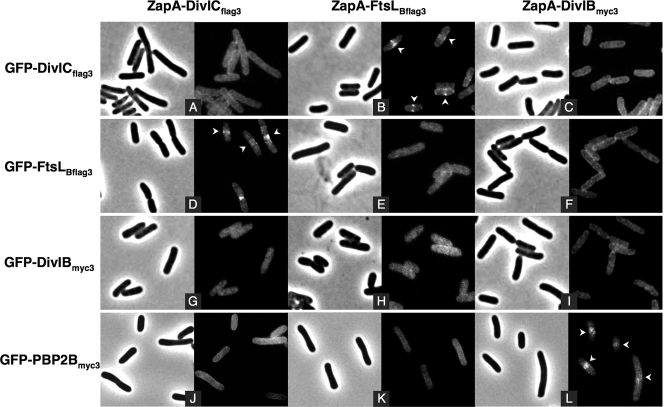
FIG. 3.
Interaction assays with DivICflag3, FtsLBflag3, DivIBmyc3, and PBP 2Bmyc3 fused to GFP and ZapA using the E. coli artificial septal targeting. Fluorescence microscopy images show subcellular localization of DivICflag3, FtsLBflag3, DivIBmyc3, and PBP 2Bmyc3 GFP fusions when coexpressed with DivICflag3, FtsLBflag3, or DivIBmyc3 ZapA fusions. The phase-contrast images corresponding to the same field as the fluorescence images are shown. Strains (from CR209 to CR211 [A to C], CR213 to CR215 [D to F], CR217 to CR219 [G to I], and CR374 to CR376 [J to L], Table 1) were grown at 30°C, and fusions were induced during 30 min with 20 μM IPTG. Samples were then prepared for microscopy as described in Materials and Methods. Arrows indicate mid-cell fluorescence.

