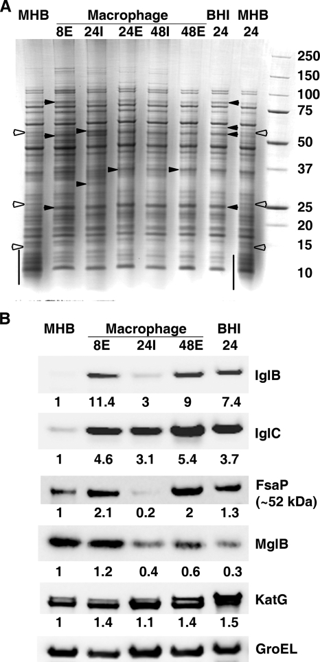
FIG. 3.
BHI-grown Francisella organisms most closely resemble extracellular, Mφ-grown bacteria. (A) MHB-grown F. tularensis LVS (lane MHB) was used to inoculate fresh MHB and BHI broths and to infect a murine Mφ cell line. At the indicated times (h), bacteria were harvested from the culture media and the extracellular (E) or intracellular (I) compartment of the Mφ cultures. Lysates (10 μg/lane) of each were resolved by SDS-PAGE, followed by staining with Coomassie blue or transfer to nitrocellulose for Western blot analysis. (A) Arrowheads and lines are as described in the legend to Fig. 1. (B) A single membrane was probed (as in Fig. 1) with a cocktail of MAbs against IglB and IglC, followed sequentially by sera against FsaP, MglB, KatG, and GroEL. Numerical values below each blot are GroEL-normalized levels of induction over those observed with MHB-grown cells.

