
FIG. 3.
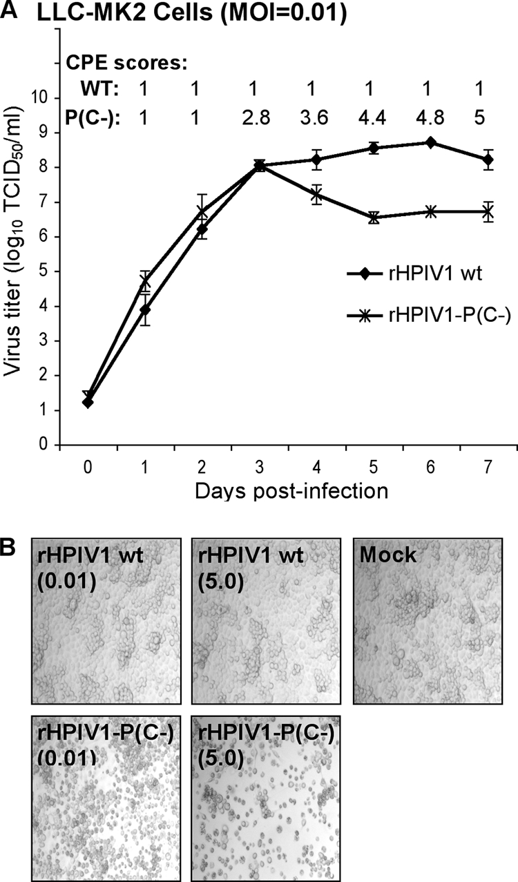
Comparison of the replication of wt rHPIV1 and rHPIV1-P(C−) in vitro. (A) Multicycle replication in LLC-MK2 cells infected at an MOI of 0.01 TCID50/cell. On days 0 to 7 p.i., the overlying medium was harvested for virus titration, shown as the means ± SE of the results for three replicate cultures. On days 1 to 7 p.i., the cell monolayers were monitored for CPE and assigned a score of 1 to 5 according to the extent of CPE (Materials and Methods), shown as the means of the results for the three replicate cultures. (B) LLC-MK2 cells were mock-infected or infected with wt rHPIV1 or rHPIV1-P(C−) at an MOI of 0.01 or 5 TCID50/cell, as indicated in parentheses below the virus names. Photomicrographs taken at 72 h p.i. show increased CPE in the rHPIV1-P(C−)-infected cultures (magnification, ×10).