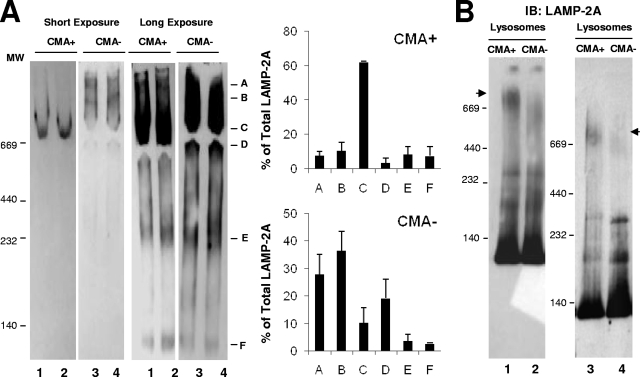
FIG. 4.
The organization of LAMP-2A into protein complexes is different in lysosomes with different CMA activity. (A) Lysosomes with high (CMA+) and low (CMA−) CMA activity isolated from the livers of 48-h-starved rats were subjected to native gel electrophoresis and immunoblotted for LAMP-2A. Left, two different exposures of a representative immunoblot. Right, mean values + SE of the distribution of LAMP-2A in the different complexes were calculated by the densitometric quantification of three to four immunoblots as the ones shown. Values are expressed as percentages of the total amount of LAMP-2A at the lysosomal membrane. (B) The same groups of lysosomes were subjected to BNE and immunoblotting (IB) of LAMP-2A. The immunoblot on the right shows a gel run for a longer time to increase the resolution of the intermediate complexes. Arrowheads indicate the approximately 700-kDa complex.

