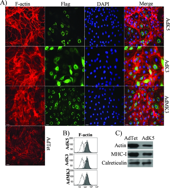
FIG. 6.
Dysregulation of the actin cytoskeleton by K5. (A) F-actin staining (red) in the presence of K5, KSHV K3, or MHV68 K3 (green). E-DMVECs were transduced with AdTet alone or together AdK5, AdK3, or an Ad vector expressing MHV68 K3 (AdMK3) for 24 h and then subjected to IFA. Viral proteins were visualized with fluorescein isothiocyanate-conjugated anti-FLAG antibody, whereas F-actin was stained with phalloidin. (B) Intracellular flow cytometry analysis for F-actin in E-DMVECs transduced with AdTet alone (shading) or together with the indicated Ad constructs (solid line). Background staining by the secondary antibody is indicated by dashed lines. (C) The total amount of actin in E-DMVECs transduced with AdK5 and that in cells transduced with AdTet were immunoblotted using anti-F-actin antibody from Abcam. MHC-I was detected with a polyclonal antiserum (K455).

