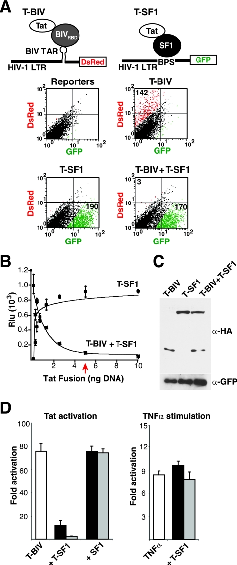
FIG. 1.
A potent dominant negative Tat inhibitor identified in a reporter assay. (A) The top illustrations represent a schematic for a dual-reporter fluorescence assay in which T-BIV is used to activate an HIV-1 LTR-BTAR-DsRed reporter. The T-SF1 fusion protein is used to activate an HIV-1 LTR-BPS-GFP reporter (Tables 1 and 2). The graphs shows results for HeLa cells cotransfected with both reporters and T-BIV and/or T-SF1 activators as indicated and sorted by flow cytometry. Expression of GFP is shown in green, and expression ofDsRed is shown in red. Numbers in each quadrant represent activation (n-fold). (B) Dose-response curves of T-SF1 activation on an HIV-1 LTR-BPS-FFL reporter and T-SF1-mediated inhibition of T-BIV activity on an HIV-1 LTR-BTAR-FFL reporter. Rlu, relative luciferase units. The arrow indicates stoichiometric DNA concentrations of inhibitor and activator. (C) Western blot of HeLa cells cotransfected with HA-tagged versions of the T-BIV activator and/or the T-SF1 inhibitor along with a GFP expressor to normalize for transfection efficiency. α, anti. (D) The left panel shows results for HeLa cells transiently transfected with the HIV-1 LTR-BTAR-FFL reporter plasmid and different ratios of T-BIV activator to T-SF1 or unfused SF1 plasmids (1:0.2, black bars; 1:1, gray bars). The right panel shows results for HeLa cells transiently transfected with the HIV-1 LTR-BTAR-FFL reporter plasmid and two amounts of T-SF1 plasmid (1 ng, black bars; 5 ng, gray bars), and transcription was stimulated 16 h posttransfection by incubation with TNF-α (10 ng/ml) for 4 h.

