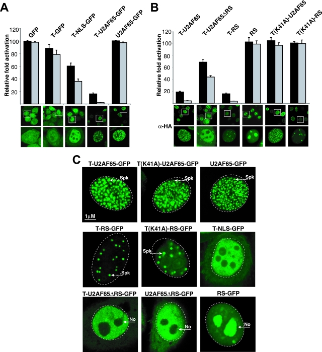
FIG. 4.
Domains that contribute to inhibition and subcellular localization. (A) HeLa cells were transiently cotransfected with an HIV-1 LTR-RREIIB-FFL reporter plasmid, T-Rev activator, and various inhibitors at 1:0.2 (black bars) or 1:1 (gray bars) ratios of activator to inhibitor. Activation levels are plotted as relative levels of activation, normalized to the level of activation without inhibitor (85-fold). Confocal images of each GFP-tagged fusion, including images with magnification of ×3 (lower images, representing the boxed cells shown in the upper images) to highlight the subcellular compartments, are shown below the graph. T-NLS-GFP contains the eight-amino-acid NLS (PPKKKRKV) of simian virus 40 T-Ag (Table 2). (B) Relative activities of T-U2AF65 deletion mutants and Tat AD variants, as determined in experiments whose results are shown in panel A. Corresponding confocal anti-HA (α-HA) immunofluorescence images are shown. T-U2AF65ΔRS-HA has a deletion of the first 90 amino acids of U2AF65, and T-RS contains only residues 2 to 73. (C) Subnuclear localization of active and inactive Tat inhibitors. HeLa cells were transfected with pEGFP-N3 plasmids expressing GFP fused to U2AF65, T-U2AF65 (dominant negative), T(K41A)-U2AF65 (inactive dominant negative), RS (U2AF65 RS domain only), T-RS (active dominant negative), T(K41A)-RS (inactive dominant negative), U2AF65ΔRS, and T-U2AF65ΔRS. Spk indicates the position of nuclear speckles; No indicates nucleoli.

