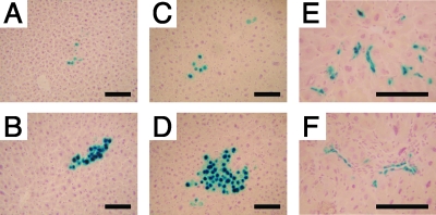
FIG. 4.
X-Gal-positive hepatocyte clusters and nonparenchymal cell clusters in livers transduced with vector AAV8-EF1α-nlslacZ at birth. Representative photomicrographs of X-Gal-positive hepatocyte clusters (A to D) and nonparenchymal cell clusters (E and F) in liver sections are shown. (A) Wild-type liver with no hepatectomy; (B) wild-type liver after hepatectomy; (C) 44Bri liver with no hepatectomy; (D, E, and F) 44Bri liver after hepatectomy. These nonparenchymal cells appear to be endothelial cells, although the exact identities of these cells have yet to be determined. Each section was stained with X-Gal and counterstained with hematoxylin. Scale bars represent 100 μm.

