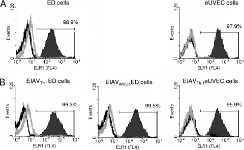
FIG. 1.
ELR1 remains on the surface of EIAV-infected cells. ED cells and eUVECs (A) and chronically EIAV-infected cells (B) were immunostained for surface expression of ELR1 using rabbit polyclonal antisera against the ectodomain of ELR1. EIAV-infected populations were greater than 90% positive for EIAV antigens. Allophycocyanin-conjugated goat-anti-rabbit was used as a secondary antibody. Flow cytometry of live, stained cells was used to determine the percentage of the population that was positive for ELR1 (solid area). Secondary antisera alone (black line) as well as rabbit control sera (gray line) were used as negative controls. A representative flow experiment is shown. The experiments were performed three independent times.

