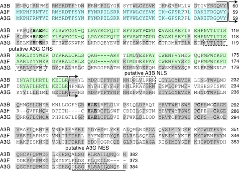
FIG. 1.
Alignment of A3B, A3F, and A3G amino acid sequences, highlighting regions of concentrated identity. Blue and green shading indicates regions of concentrated identity between A3G and A3F and between A3B and A3F, respectively. Gray-shaded residues are identical between at least two of the proteins. The crossing arrows indicate the junctions of chimeric proteins. Dashed boxes outline the putative CRS and NES of A3G and the putative NLS of A3B. The histidine, glutamic acid, and cysteine residues of the two zinc-coordinating motifs are indicated in boldface. The amino acids deleted in A3G1-369 are underlined. Additional details can be found in the text.

