
FIG. 2.
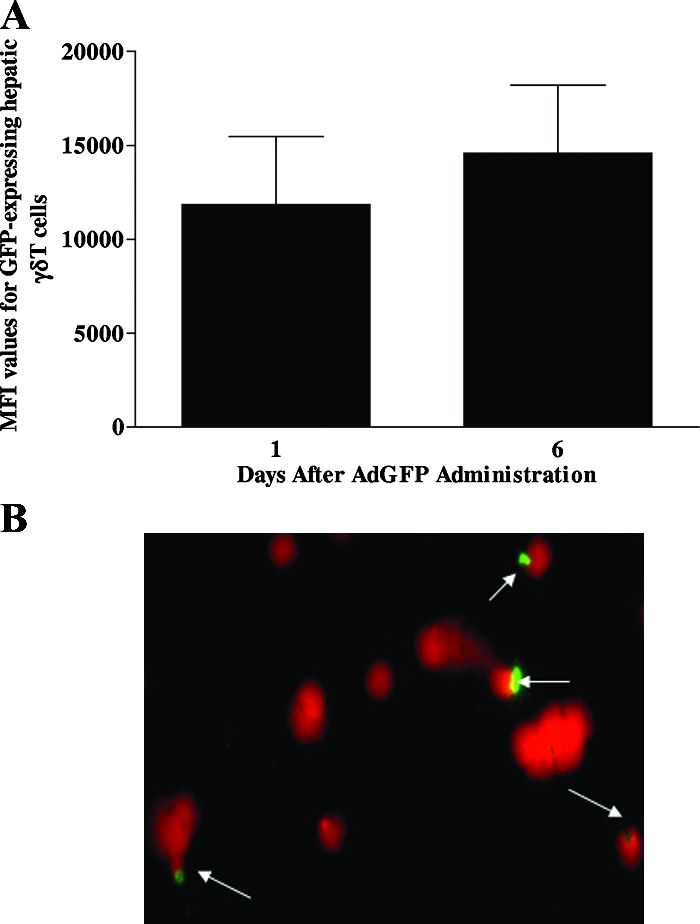
Ad interacts with γδT cells. (A) Male C57BL/6 mice were injected with AdGFP, and isolated hepatic fluorochrome-labeled γδT cells were screened for the presence of GFP by flow cytometry. The median fluorescence intensity (MFI) values for GFP-positive hepatic γδT cells are shown. Data are reported as means ± SEM (n = 4 mice per group). (B) To determine if Ad could interact with γδT cells, splenocytes were isolated from naive C57BL/6 mice and γδT cells were purified using a mouse γδT-cell isolation kit. Purified splenic γδT cells were then incubated in vitro with GFP-labeled Ad (104 virus particles) for 60 min. A fluorescence microscopic technique was used to evaluate Ad interaction with splenic γδT cells. Ad-GFP is shown in green, whereas γδT-cell nuclei counterstained with propidium iodide are shown in red.