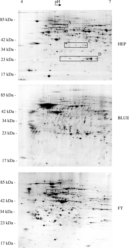
FIG. 2.
Scans of 2D electrophoretic gels representing the two affinity-purified fractions (BLUE, cibacron fraction; HEP, heparin fraction) and the flowthrough (FT) of the ASPE separation procedure. Differing protein patterns reflect an efficient separation into three well-defined protein fractions. Boxes indicate gel regions that were analyzed in more detail (Fig. 6 and 7) for modifications of eIF-4B and SNX-9 (box A), lamin B2 (box B), 60S acidic ribosomal protein P0 (box C), Hsp27 (box D), and hnRNP K (box in panel FT).

