FIG. 3.
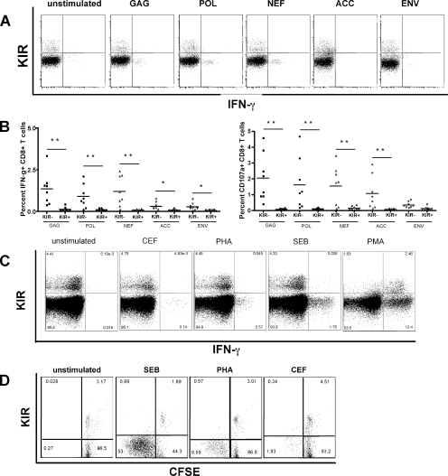
KIR expression results in reduced TCR-mediated CD8+ T-cell activation. (A) The flow plots demonstrate the reduced capacity of KIR+ CD8+ T cells from a representative HIV-positive subject to secrete cytokines following stimulation with HIV peptide pools. (B) The dot plots show the level of IFN-γ secretion (left panel) and CD107a upregulation (right panel) on KIR+ and KIR− CD8+ T cells following stimulation with peptide pools spanning individual HIV gene products. (C) Flow plots depict the ability of KIR+ and KIR− CD8+ T cells to secrete IFN-γ following stimulation via the TCR (CEF, PHA, and SEB) and PMA, which activates T cells outside the TCR in a single representative subject. (D) The diagram shows the level of CFSE dilution in KIR+ and KIR− CD8+ T cells following stimulation and demonstrates that proliferation is also impaired in KIR+ CD8+ T cells.

