Figure 2.
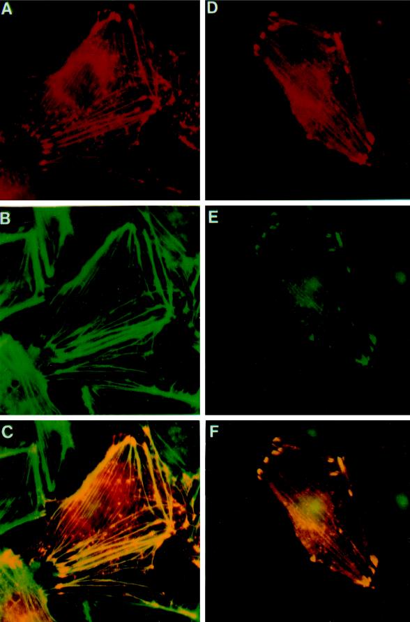
Colocalization of Ena and VASP proteins to stress fibers and focal contacts in transfected PtK2 cells. PtK2 cells were transiently transfected with pCMV/ena (A–C) or pCMV/ena and pVSV-VASP (D–F) and processed for double-label immunofluorescence microscopy. Ena was detected with rabbit polyclonal antibodies (A and D), VASP with a monoclonal mouse anti-VSV antibody (E), and F-actin by fluorescein-labeled phalloidin (B). A merge of A and B is shown in C, and a merge of D and E is shown in F. Fluorescent staining was by TRITC for Ena and DTAF for VASP. Ena expressed alone is localized either in focal contacts and stress fibers or is also variably found in spot-like structures, which also stain for F-actin (A–C). Coexpression of Ena and human VASP results in an identical localization of the two proteins in focal contacts and stress fibers (D–F). Controls show no cross-reactivity of the anti-Ena antibodies with VASP. Nuclear fluorescence is nonspecific background because of the secondary antibody.

