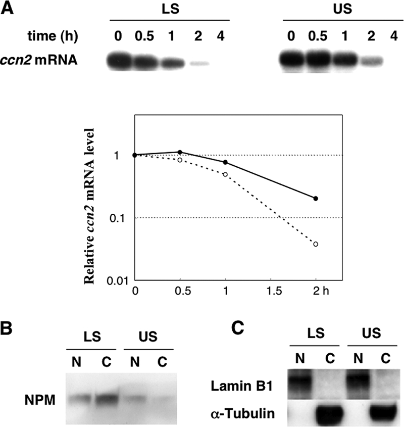
FIG. 4.

Relationship between intracellular ccn2 mRNA stability and subcellular distribution of NPM in chondrocytes. (A) Degradation profiles of ccn2 mRNA in LS and US cells. RNA synthesis in LS or US cells was arrested by actinomycin D, and a time-course study of the fate of the remaining ccn2 mRNA was analyzed by Northern blotting analysis (top panels). The level of 28S rRNA remained unchanged through the experimental time course (data not shown). The numbers (hours) indicate the time periods after transcriptional arrest was initiated. The signal intensities of the autoradiograms were quantitatively analyzed and plotted compared to the values at time zero (bottom panel). The broken line with open circles and solid line with closed circles denote the data obtained with LS and US cells, respectively. (B) Nucleocytoplasmic distribution of NPM in LS and US cells. Proteins from the nuclear extract (N) or cytoplasmic extract (C) from cells comprising the same amount of DNA were subjected to 12.5% SDS-PAGE and blotted onto a PVDF membrane. The blot was then incubated with anti-NPM antibody and then with secondary antibodies for visualization of signals, as described in Materials and Methods. (C) Successful fractionation confirmed by Western blotting of each fraction (10 μg protein) with anti-lamin B1 (nuclear marker) or anti-α-tubulin (a cytoplasmic marker) antibody.
