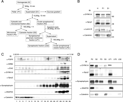
FIG. 6.
DYRK1A and Spry2 associate with the plasma membranes of synaptic terminals. (A) Schematic representation of the subcellular fractionation procedures used for the analyses in panels B to D. Centrifugation steps are indicated with a semicircular arrow. Full details are provided in Materials and Methods. (B) Homogenates from mouse brain were obtained as described in Materials and Methods. Equal volumes of the homogenate (H), the nuclear pellet (P1), and the postnuclear fraction (S1) were analyzed by Western blotting with the indicated antibodies. (C) The postnuclear fraction (S1) from mouse brain homogenates was fractionated by centrifugation in a discontinuous sucrose gradient. Equal volumes of the first 18 alternate fractions were analyzed by Western blotting with specific antibodies, as indicated. (D) The postnuclear fraction (S1) from mouse brain homogenates was fractionated by differential centrifugation. Equal volumes of the fractions obtained were analyzed by Western blotting with specific antibodies, as indicated. P2, crude synaptosomal fraction; S2, cytosolic and microsomal fraction; P3, microsomal fraction; S3, soluble cytosolic fraction; LP1, synaptosomal plasmatic membrane fraction; LP2, crude synaptosomal vesicle fraction; LS2, synaptosolic fraction.

